Radial head fracture
From Wikipedia, the free encyclopedia
| Radial head fracture | |
|---|---|
 | |
| Radial head fracture (red arrow) with posterior and anterior sail sign (blue arrows) | |
| Specialty | Orthopedics |
| Symptoms | Pain or tenderness over the radial head; bruising; swelling; limited range of motion. |
| Causes | Fall on an outstretched arm |
| Diagnostic method | Based on clinical symptoms and medical imaging |
| Treatment | Varies according to severity of injury but may include: immobilization followed by range of motion exercises; joint aspiration with mobilization; surgical correction |
Radial head fractures are a common type of elbow fracture that typically occurs after a fall on an outstretched arm.[1] They account for approximately one third of all elbow fractures and are frequently associated with other injuries of the elbow.[2][3] Radial head fractures are diagnosed by a clinical assessment and medical imaging.[2][4] A radial head fracture is treated according to the severity of the injury and its Mason-Johnston classification. Treatment may be surgical or nonsurgical. Stable isolated fractures typically have excellent outcomes.[5] Unstable fractures with other associated injuries have varying outcomes. Common adverse outcomes include stiffness, pain, poor bone healing, and hardware complications.[6]
Diagnosis and classification
Radial head fractures are diagnosed from a clinical assessment and diagnostic imaging.[7] Symptoms may include pain or tenderness at the radial head, bruising, swelling, and a limited range of motion of the injured elbow.[2] Diagnostic imaging may include ultrasound, plain radiography (x-ray imaging), Computed tomography scan (CT), and magnetic resonance imaging (MRI).[2][4] A fat pad sign may be present on diagnostic imaging and may indicate a radial head fracture.[5]
A diagnosed radial head fracture can be classified according to the Mason-Johnston system.[3]
| Type | Description |
|---|---|
| 1 | Non-displaced fracture |
| 2 | Minimal displacement with angulation or impression (>2mm) |
| 3 | Comminuted fracture with dislocation |
| 4 | Radial head fracture with dislocation of the elbow |
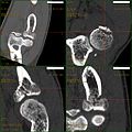 CT scan showing a radial head fracture
 Radial head fracture seen on 3D CT reconstruction


Treatment
Radial head fracture treatment is informed by the Mason-Johnston classification, patient symptoms, and fracture stability. An unstable fracture will involve fracture displacement, fractures to adjacent structures and injury to other associated soft tissues. A stable type 1 radial head fracture is typically managed with conservative measures including joint aspiration, immobilization in a sling for a few days and followed by early range of motion exercises.[2][6] If range of motion is still limited after joint aspiration it may indicate a mechanical block which is treated surgically.[5] Stable type 2 radial head fractures may be treated as a type 1 if the displacement is minimal. Unstable type 2 - 4 fractures generally warrant surgery. Surgical correction can include fracture fragment excision, radial head reconstruction, open reduction and internal fixation, and radial head excision with artificial replacement.[6] Associated structures that were damaged during the injury may also need to be repaired.
Rehabilitation exercises are recommended and tailored to fracture and treatment type. It is recommended to wait 6 weeks before resuming load bearing with a stable type 1 fracture and 10-12 weeks following surgery for unstable type 2-4 fractures.[8]