T7 DNA polymerase
From Wikipedia, the free encyclopedia
| DNA-directed DNA polymerase | |||||||
|---|---|---|---|---|---|---|---|
 Figure 1. Crystal structure of T7 DNA replication complex. Rendered from PDB 1T7P. | |||||||
| Identifiers | |||||||
| Organism | |||||||
| Symbol | 5 | ||||||
| CAS number | 9012-90-2 | ||||||
| UniProt | P00581 | ||||||
| Other data | |||||||
| EC number | 2.7.7.7 | ||||||
| |||||||
T7 DNA polymerase is an enzyme used during the DNA replication of the T7 bacteriophage. During this process, the DNA polymerase “reads” existing DNA strands and creates two new strands that match the existing ones. The T7 DNA polymerase requires a host factor, E. coli thioredoxin,[1] in order to carry out its function. This helps stabilize the binding of the necessary protein to the primer-template to improve processivity by more than 100-fold, which is a feature unique to this enzyme.[2] It is a member of the Family A DNA polymerases, which include E. coli DNA polymerase I and Taq DNA polymerase.
This polymerase has various applications in site-directed mutagenesis[3] as well as a high-fidelity enzyme suitable for PCR.[4] It has also served as the precursor to Sequenase,[5] an engineered-enzyme optimized for DNA sequencing.[6]
Phosphoryl transfer
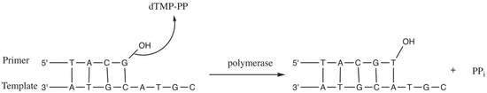
Figure 2. Nucleotidyl transfer by DNA polymerase.
T7 DNA polymerase catalyzes the phosphoryl transfer [7] during DNA replication of the T7 phage. As shown in Figure 2, the 3’ hydroxyl group of a primer acts as a nucleophile and attacks the phosphodiester bond of nucleoside 5’-triphosphate (dTMP-PP). This reaction adds a nucleoside monophosphate into DNA and releases a pyrophosphate (PPi). Generally, the reaction is metal-dependent and cations such as Mg2+ are often present in the enzyme active site.[7]
For T7 DNA polymerase, the fingers, palm and thumb (Figure 1) position the primer-template so that the 3’-end of the primer strand is positioned next to the nucleotide-binding site (located at the intersection of the fingers and thumb).[8] The base pair formed between the nucleotide and the template base fits nicely into a groove between the fingers and the 3’-end of the primer.[8] Two Mg2+ ions form an octahedral coordinate network with oxygen ligand and also bring the reactive primer hydroxyl and the nucleotide α-phosphate close together, thereby lowering the entropic cost of nucleophilic addition.[8] The rate-limiting step in the catalytic cycle occurs after the nucleoside triphosphate binds and before it is incorporated into the DNA (corresponding to the closure of the fingers subdomain around the DNA and nucleotide).[8]

Role of Mg2+ ions and amino acid residues in the active site
The amino acids present in the active site assist in creating a stabilizing environment for the reaction to proceed. Amino acids such as Lys522, Tyr526, His506 and Arg518 act as hydrogen bond donors. The backbone carbonyl of Ala476, Asp475 and Asp654 form coordinate bonds with the Mg2+ ions.
Asp475 and Asp654 form a bridge with the Mg2+ cations to orient them properly. The Mg2+ ion on the right (Figure 3) interacts with negatively charged oxygens of the alpha(α), beta(β) and gamma(γ) phosphates to align the scissile bond for the primer to attack.[8] Even if there is no general base within the active site to deprotonate the primer hydroxyl, the lowered pka of the metal-bound hydroxyl favors the formation of the 3’-hydroxide nucleophile.[8] Metal ions and Lys522 contact non-bridging oxygens on the α-phosphate to stabilize the negative charge developing on the α-phosphorus during bond formation with the nucleophile.
Moreover, the Lys522 sidechain also moves to neutralize the negatively charged pyrophosphate group. Tyr526, His506, Arg518 side chains and the oxygen from the backbone carbonyl group of Ala476 take part in the hydrogen bond network and assist in aligning the substrate for phosphoryl transfer.[8]
Accessory proteins

While phage T7 mediates DNA replication in very similar manner to higher organisms, T7 system is generally simpler compared to other replication systems. In addition to T7 DNA polymerase (also known as gp5), T7 replisome requires only four accessory proteins for proper function: host thioredoxin, gp4, gp2.5, and gp1.7.
Host thioredoxin
T7 polymerase by itself has a very low processivity. It dissociates from the primer-template after incorporating about 15 nucleotides. Upon infection of the host, T7 polymerase binds to host thioredoxin in 1:1 ratio. The hydrophobic interaction between thioredoxin and T7 polymerase helps to stabilize the binding of T7 polymerase to primer-template. In addition, the binding of thioredoxin increases T7 polymerase processivity to nearly 80-fold.[9] The precise mechanism for how the thioredoxin-T7 polymerase complex is able to achieve such increase in processivity is still unknown. Binding of thioredoxin exposes a large number of basic amino acid residues in the thumb region of T7 polymerase. Several studies suggest that the electrostatic interaction between these positively charged basic residues with the negatively charged phosphate backbone of DNA and other accessory proteins is responsible for increased processivity in gp5/thioredoxin complex.[9][10][11]
gp4
gp4 is a hexameric protein containing two functional domains: helicase domain and primase domain. The helicase domain unwinds double-stranded DNA to provide template for replication. The C-terminal tail of helicase domain contains several negatively charged acidic residues which make contact with the exposed basic residue of T7 polymerase/thioredoxin. These interactions help to load T7 polymerase/thioredoxin complex onto replication fork. The primase domain catalyzes the synthesis of short oligoribonucleotides. These oligoribonucleotides, called primers, are complementary to the template strand and used to initiate DNA replication. In T7 system, primase domain of one subunit interacts with primase domain of adjacent subunit. This interaction between primase domains acts as a brake to stop helicase when needed, which ensure the leading stand synthesis in-pace with lagging stand synthesis.[11]
gp2.5
| Single-stranded DNA-binding protein | |||||||
|---|---|---|---|---|---|---|---|
| Identifiers | |||||||
| Organism | |||||||
| Symbol | 2.5 | ||||||
| UniProt | P03696 | ||||||
| |||||||
gp2.5 has similar function to single-stranded DNA binding protein. gp2.5 protects single-stranded DNA produced during replication and coordinates synthesis of leading and lagging strands through interaction between its acidic C-terminal tail and gp5/thioredoxin.[11]
gp1.7
| Nucleotide kinase | |||||||
|---|---|---|---|---|---|---|---|
| Identifiers | |||||||
| Organism | |||||||
| Symbol | 1.7 | ||||||
| UniProt | P03781 | ||||||
| |||||||
gp1.7 is a nucleoside monophosphate kinase, which catalyzes the conversion of deoxynucleoside 5'-monophosphates to di and triphosphate nucleotides, which accounts for the sensitivity of T7 polymerase to dideoxynucleotides (see Sequenase below).[11]